
Functional and Systems Biology
A Molecular View of Cells, Proteins, and Viruses
Cryo-electron microscopy’s powerful imaging tools for structural biology research

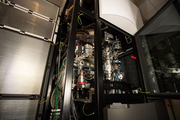
The Environmental Molecular Sciences Laboratory's cryo-focused ion beam-scanning electron microscope and a dedicated cryo-transmission electron microscope enable high-resolution structural determination of biological structures. Photo by Andrea Starr | Pacific Northwest National Laboratory)
* Updated Aug. 29, 2022
Meet cryo-electron microscopy.
This herculean technology uses electrons with velocity approaching the speed of light and keeps samples at cryogenic temperatures of −190 degrees Celsius.
And, when it comes to structural biology research, cryo-electron microscopy, or cryo-EM, has emerged as the clear front runner for high-resolution imaging focused on the molecular structure of proteins, cells, and viruses.
A powerful suite of cryo-EM instrumentation, which includes cryo-focused ion beam-scanning electron microscopy and a dedicated cryo-transmission electron microscope, is now available for use at the Environmental Molecular Sciences Laboratory (EMSL), a Department of Energy, Office of Science user facility on the Pacific Northwest National Laboratory campus in Richland, Washington.
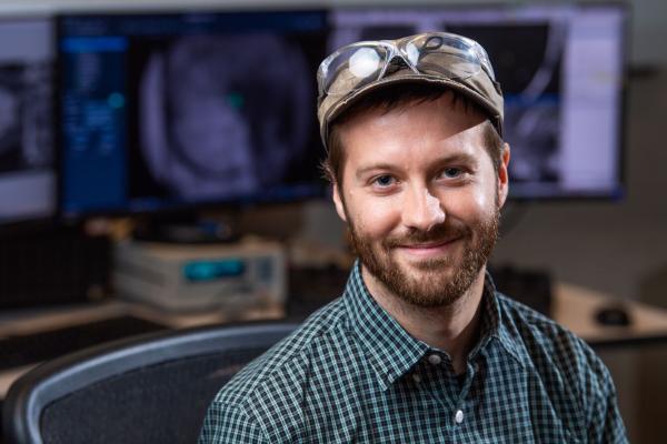
“The EMSL cryo-EM facilities offer state-of-the-art instrumentation enabling high-resolution structural determination of biological structures from a range of sample types. From target genes, to purified proteins, crystals, or whole cells, our sample preparation pipeline can prepare a wide range of diverse samples for high-resolution microscopy,” says Trevor Moser, an EMSL chemist whose research uses cryo-electron microscopy to study protein structures.
EMSL is holding a free webinar at noon Pacific daylight time on Aug. 31 to share how users can access these cryo-EM resources for structural biology research.
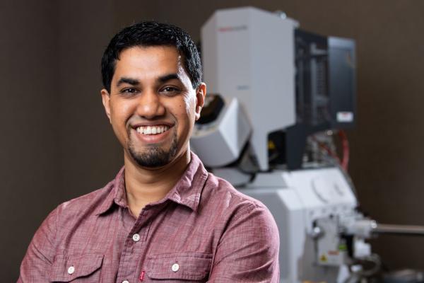
Moser will provide an overview of how the instrumentation suite is used to visualize in situ cell ultrastructure at nanometer-to-angstrom resolution. EMSL chemist Amar Parvate will explain how EMSL’s cryogenic transmission electron microscope (cryo-TEM) is used to determine the structure of proteins and protein–DNA complexes at near-atomic resolution. EMSL chemist James Evans will share how cryo-TEM applies micro-electron diffraction to study small molecules and proteins in 3D crystals.
Cryo-focused ion beam-scanning electron microscopy
In EMSL’s Quiet Wing is the Aquilos 2, a cryo-focused ion beam-scanning electron microscope (cryo-FIB-SEM) and one half of the user facility’s suite of cryo-EM instrumentation. The Quiet Wing was engineered to protect high-resolution microscopes like Aquilos from acoustic noise, vibrations, and stray electromagnetic field sources.
The Aquilos is a semi-automated instrument that is used primarily for preparing samples for high-resolution imaging in EMSL’s cryo-TEM, a Krios G3i. The Aquilos’ unique function is its ability to thin samples that are conventionally too thick to image natively with cryo-TEM into samples that are 200 to 300 nanometers thick using a gallium ion beam. By first thinning the bulk sample into thin lamellas in the Aquilos, the Krios cryo-TEM can then be used to visualize the architecture of cells using electron tomography or crystals using micro-electron diffraction.
“The cool thing about Aquilos is that it greatly expands the type of samples we can work with,” says Moser, who notes that Aquilos has been used to prep samples for human health, purified proteins, and plant research.
Moser and EMSL research staff have two workflows that are used for lamellas—a cryogenic batch lamella generation workflow, and a site-selective method where a region is removed and then attached to a TEM grid to be thinned and imaged. In addition to sample preparation, the Aquilos may be used to generate 3D volumes of clumps of cells or tissues at nanometer resolution with a technique known as “slice and view” imaging.
Cryo-transmission electron microscopy
In Greek mythology, Krios is the Titan God of Constellations. At EMSL, Krios is the masterful technology that gives researchers a nanoscale view of cells, proteins, and tissues.
"The Krios—with all the imaging modalities and related software—makes studying [the] biology scale agnostic,” says Parvate. “The Krios routinely allows collection of ginormous datasets, which had not been possible a decade ago. Now structural biology is not limited by [the] amount of data collected."
The Krios cryo-TEM offers three workflows—cryo-EM single particle analysis, micro-electron diffraction, and cryo-electron tomography—to study protein structures at the molecular level.
The Krios is used to conduct single particle analysis, trapping protein samples in vitreous ice and then collecting thousands of two-dimensional structural images of the purified proteins. By combining and aligning the signals of tens to hundreds of thousands of individual particles, the 3D structure of the protein sample may be determined.
With micro-electron diffraction, the Krios pulls three-dimensional atomic information from individual nanocrystals to obtain structural data about molecules and proteins. Micro-electron diffraction can determine the structures of small molecules, peptides, and small proteins that are too small to observe with single particle analysis and cryo-electron tomography, assuming they can be crystalized.
Cryo-electron tomography is a technique that takes two-dimensional images through incremental tilts from a lamella or thin sample and reconstructs the resulting tilt series images into a three-dimensional volume. This data can be used to investigate changes in cellular ultrastructure, and in some cases may be used to solve the structures of large protein complexes in their native cellular environments.
Combined, these techniques each allow access to a wide range of biological structures, from small molecules to tissues, and allow for a large degree of sample flexibility. EMSL is equipped to accept user samples in a range of different formats, and staff is available to work with users to identify the best route for generating structural data for their projects.
Structural biology samples for cryo-electron microscopy
Cryo-electron microscopy has a particular set of requirements for sample success. Topping the list is that the sample must be vitrified to preserve its ultrastructure. In doing so, the cell environments and culture conditions can be preserved until they are plunge-frozen.
Structural biology samples should include the following:
-
Isolated/purified protein complexes
-
Nanocrystals and microcrystals
-
Host-viral/phage interactions
-
Plant–microbe–fungi interactions under different environmental conditions
-
Biosystems with normal or environmentally stressed conditions
Proposal opportunities
The suite of cryo-TEM instrumentation is accessible through two proposal mechanisms.
EMSL’s User Program offers proposal opportunities throughout the year, enabling the use of cryo-TEM with other capabilities.
Proposals that will use only cryo-EM/ET or micro-electron diffraction are also accepted, but undergo an accelerated review without a specific proposal due date. Evans is the contact for these proposals.
Questions about applying cryo-EM to user research can be addressed at the Aug. 31 webinar or by contacting Moser, Parvate, or Evans.